


Papillomas on the neck are one of the manifestations of an infectious disease caused by the human papillomavirus.They belong to benign skin formations.
Causes of papillomas on the neck
There is an etiological reason why papillomas begin to grow on the neck or in any other area of the human body - infection with the human papillomavirus (papillomavirus, HPV), which represents the Papovaviridae family.There are more than 100 serotypes of this pathogenic agent, each of them is responsible for the appearance of a different clinical picture of the disease (papilloma, condylomas, warts - these concepts are synonymous, different names are associated with the peculiarities of localization in a certain area).
The main routes of transmission are through household contact and sexual contact (condylomas of the perianal region).The virus can only penetrate the skin in the presence of microdamage or open wounds;in other cases, it cannot penetrate the skin's protective barrier.
Pathogen Information
- It has a high prevalence regardless of gender (however, it occurs in women a little more frequently than in men), age or region (according to some data, 2/3 of the planet is infected with this virus).
- It contains double-stranded, circular, twisted DNA that can be integrated into the human genome.
- Infection with some strains is associated with a high carcinogenic risk, especially in the case of permanent damage.Papillomas on the neck are caused by non-oncogenic strains of the virus.
- During the division process, the virus goes through two main steps.In the first phase, it appears in the episomal (free) form and in the same period the main division of the viral particle occurs.This phase is reversible (long-term remission occurs after treatment).In the second stage - integrative - the virus is implanted into the cell genome (the first step towards cell degeneration and the formation of a malignant neoplasm).The first phase is transient and passes relatively quickly, and the second is latent and explains the existence of carriers.
- The basal layer of the epidermis is affected, where virus replication occurs.In the remaining layers, the pathogen can persist but not divide.Since the virus is in the germinal layer, as it grows, the normal differentiation of cells in all layers of this area is disturbed, the disturbances are especially strong at the level of the spinous layer.
- It has a tendency to long-term asymptomatic carriage in the body (from several months to a year).It is rarely possible to identify the specific moment of infection - that is why treatment begins during the period of intense clinical manifestations, and not at the first vague signs.
- To prevent infection, bivalent and quadrivalent vaccines are used, which are especially effective against the most oncogenic strains 16 and 18.
Predisposing factors
- Failure to comply with hygiene rules.Since the virus is able to maintain vital activity in the external environment for a long time, it is necessary to carefully observe the rules of personal hygiene when visiting public places (swimming pool, sauna, gym).
- Traumatic skin injuries.Microcracks or scratches in the skin (for example, caused by rubbing the neck with a shirt collar) are sufficient for the virus to penetrate.
- Immune system dysfunction.In immunodeficiencies of any origin, favorable conditions arise for the development of any infections.For example, frequent colds and infectious diseases lead to a weakening of immunity and the appearance of papillomas on the skin.
- Self-infection by scratching the skin.
- Systematic lifestyle disorders (stress, lack of physical activity, unhealthy diet).These factors affect the functioning of all metabolic processes in the body and lead to a decrease in the skin's barrier function.
- Environmental factors that influence the reduction of the body's defenses (hypothermia, excessive exposure to ultraviolet rays).
External manifestations of the disease
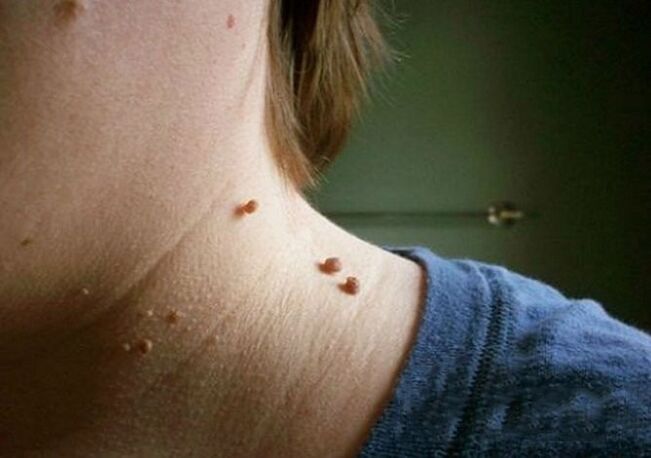
The cervical papillomas in the photo look like this:
- The growth is usually located on a wide base and protrudes significantly above the surface of the skin.Less commonly, the base of the papilloma is represented by a thin rod (in this case, the formation is suspended).In the second option, the risk of injury is much greater.
- The boundaries of education are soft and clear.
- The color does not differ from the surrounding skin.In rare cases, the surrounding tissue may be slightly paler or darker.
- The surface is usually flat and smooth.Sometimes growths are possible in the upper part of the papilloma, which makes its surface ribbed.
- The diameter varies greatly - from 1-3 mm to several centimeters (small diameter papillomas are more common).
- Location in any region of the neck (back, sides, front).Sometimes a face is involved.
As a rule, there are many lesions located along the skin folds.
In very rare cases, papillomas on the neck can become malignant, that is, degenerate into a skin tumor.This can occur as a result of infection with an oncogenic strain of HPV.
Signs that may indicate malignant degeneration are the following:
- color change and heterogeneity (polymorphism);
- edge change (blurring, loss of clarity);
- the appearance of asymmetry (when drawing a line through the conditional middle of the formation, it is impossible to obtain two equal halves);
- intensive growth;
- bleeding or ulceration (nonspecific sign, as it is also typical of simple trauma from neoplasia);
- itching, burning, peeling;
- projections are formed (small daughter formations around the central one).
The occurrence of such signs does not necessarily mean degeneration of the papilloma, but it does mean that it is necessary to consult a doctor and carry out a differential diagnosis, finding out whether it is a regular inflamed mole or skin cancer.
How to get rid of papillomas on the neck
Treatment of papillomas on the neck is carried out only comprehensively, with a simultaneous effect on the pathological focus on the skin and the pathogen itself in the blood.
You can fight in several ways:
| Method |
Description |
| Medication methods |
The use of cytostatics and immunomodulators aims to suppress the replication of the viral agent in the affected area and reduce its concentration in the blood.Some medications (keratolytics) are applied topically directly to destroy skin growth (they cauterize and cause tissue necrosis). |
| Physical methods |
Cryodestruction, laser therapy, electrocoagulation.The goal is to eliminate papillomas on the neck and other parts of the body.These methods allow you to restore the aesthetic appearance of open areas and remove the viral reservoir - the skin tumor itself, but they do not completely remove the virus from the body. |
| Combination therapy |
It combines the two previous options and is therefore more effective. |
Treatment of papillomas with folk home remedies (celandine juice, for example) is ineffective and often dangerous;in any case, a necessary condition is consultation with a doctor.
Physical methods of destruction
It is possible to effectively reduce formations using the following physical methods:
| Method |
Description |
| Local exposure to concentrated acid solutions |
A 1.5% solution of zinc chloropropionate in 50% 2-chloropropionic acid, a combination of nitric, acetic, oxalic, lactic acids and copper nitrate trihydrate, etc. are used.The procedure is performed on an outpatient basis by a specialist (dermatovenereologist, cosmetologist) in accordance with surgical standards.The product is applied pointwise with a spatula until the color of the formation changes to a lighter color (as soon as this happens, further application must be stopped immediately).To completely cure papilloma, on average you need to do 1-2 treatments. |
| Electrocoagulation |
Using a special electric knife, a targeted excision of formations is performed without affecting the underlying tissues (there is minimal impact on healthy skin cells).The method is more convenient when the formation has a long stem and small size. |
| Cryodestruction |
The lesion is exposed to liquid nitrogen;ultra-low temperature leads to tissue necrosis.It is good to remove formations with a wide base in this way.The duration of nitrogen action is selected by a specialist (1-5 minutes).After cauterization, a burn is formed that heals in an average of 10 days. |
| Laser removal |
The most modern and delicate approach, allowing you to remove formations in prominent locations such as the neck.It has the most positive reviews.Using a light guide, the lesion is exposed for 5 seconds to 3 minutes in continuous mode.The healing period is much shorter than other methods (5-7 days).The technique is associated with minimal trauma to surrounding tissues due to the high precision of the impact. |
| Classical surgical removal (scalpel excision) |
It is used extremely rarely, only for large lesions or suspected malignancy.The reason is that the lesions are often multiple, spread across the neck, and too small for excision;Furthermore, after surgical excision, scars may remain, which in turn create a cosmetic defect. |